
This skin diagram clearly shows all the layers of skin. We will now go over the skins layers in more detail.
Epidermis Layer
As can be seen in the skin diagram, the outermost layer of the skin is called the epidermis layer. There are no blood vessels in the epidermis but its deepest layer is supplied with lymph fluid. It is thickest in the palms and on the bottom of the feet.
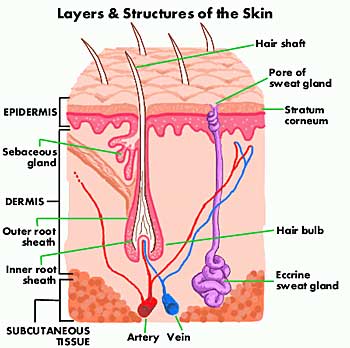
There are various layers of cells within the epidermis, the outermost of which is called the stratum corneum (or horny layer). These can be clearly seen in the diagram of skin. This surface layer is composed of twenty-five to thirty sub-layers of flattened scale-like cells. These are continually being cast off by friction and replaced by the cells of the deeper epidermal layers. This surface layer is considered the real protective layer of the skin. These cells are commonly called keratinised cells because the living matter inside the cell (termed protoplasm) is changed to a protein (keratin) that helps to give the skin its protective properties.
New skin cells are formed in the deepest layer within the epidermis. This area is called the stratum germinativum (see skin diagram). The new cells will gradually move towards the outer layers of the skin as the stratum corneum is abraded or shed. The new cells gradually change in form as they move upward to the outer layers, becoming keratinised in the process.
Dermis Or Corium Layer
The dermis is a tough and elastic layer containing white fibrous tissue interlaced with yellow elastic fibers.
As you can see in the skin diagram, many structures are embedded in the dermis including:
- blood vessels
- lymphatic capillaries and vessels
- sweat glands and their ducts
- sebaceous glands
- sensory nerve endings
- the arrectores pilorum (or arrector pilli), involuntary muscles are sometimes activated in cold weather to give “goose bumps”
- hair follicles, hair bulbs and hair roots.
Hypodermis Or Subcutaneous Layer
This is the deepest of the layers of skin, and is located on the bottom of the skin diagram. It connects or binds the dermis above it to the underlying organs. This layer is mainly composed of loose fibrous connective tissue and fat (adipose) cells interlaced with blood vessels. In females, the hypodermis is generally about 8% thicker than in males. The main functions of the hypodermis include insulation, storing of lipids, cushioning of the body and temperature regulation.
Skin Functions
- Protects the body against physical injury.
- Provides some protection for the body against numerous pathogenic microbes and chemical agents.
- Helps to restrict fluid and water loss.
- Helps to prevent excessive water absorption by imparting water resistance to the skin.
- Is involved in temperature regulation of the body.
- Is the body’s main sensory organ for temperature, pressure, touch and pain.
- Provides protection from UV light.
- Plays a key role in metabolism, including vitamin D synthesis and biotransformation of some chemicals. Lack of vitamin D can lead to soft bones and many associated problems.
Many people do not realize how much the skin does. As can be seen in the diagram of skin, your skin is a complex and important organ. It is also the largest organ in the body.
If you have any questions about our diagram of skin, please contact us. If you do not understand the functions of any layers of skin, please write to us.
More than Skin Diagram on our Skin Care page
Healthy Skin Guide Home Page